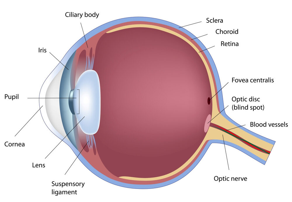
A A
  • Contact Us
  • News & Updates
  • Careers
  • For Medical Professionals
Georgia Retina
  • 888-427-3846
  • Patient Portal
  • Doctor Portal
  • Pay My Bill
  • Home
  • Contact Us
  • Doctors
    • Michael S. Jacobson, M.D.
    • Scott I. Lampert, M.D.
    • Jay Stallman, M.D., F.A.C.S.
    • Mark J. Rivellese, M.D.
    • Sean S. Koh, M.D.
    • Atul Sharma, M.D.
    • Robert Stoltz, M.D., Ph. D.
    • John J. Miller, M.D.
    • Stephanie Vanderveldt, M.D.
    • Hyung Cho, M.D.
    • Krishna Mukkamala, M.D.
    • David S. Chin Yee, M.D.
    • Paul S. Walia, M.D.
    • Yogin Patel, M.D.
    • Gregory D. Lee, M.D.
    • Ella H. Leung, M.D.
    • Rahul Komati, M.D.
  • Locations
    • Cartersville
    • Conyers
    • Cumming
    • Douglasville
    • Gainesville
    • Gwinnett (Lawrenceville)
    • Macon
    • Marietta
    • Northside
    • Peachtree City
    • Stockbridge
    • Surgery Center
    • Tucker
  • Conditions
    • Age-Related Macular Degeneration
    • Branch Retinal Vein Occlusion
    • Central Retinal Vein Occlusion
    • Central Serous Retinopathy
    • Cystoid Macular Edema
    • Diabetic Retinopathy
    • Macular Holes
    • Macular Pucker
    • Ocular Histoplasmosis
    • Retinal Detachment
    • Retinopathy of Prematurity
    • Tumors of the Eye
    • Uveitis
  • Patient Education
    • About the Eye
    • Amsler Recording Chart
    • Conditions
    • Surgical Services
    • Educational Videos
    • Useful Links
    • Testimonials
  • Clinical Trials
  • For Medical Professionals
    • Doctor Portal
  • Menu Menu

About the Eye

You can understand the way your eyes work by thinking of a camera. The white part of your eye is called the sclera. In its center is the cornea, the transparent part of the eye that covers the iris or colored part of your eye. The iris operates like a camera shutter by controlling the amount of light that enters the eye.

Located behind the iris is the eye lens. It is suspended by fibers that tighten or loosen to focus the light rays from objects outside the eye onto the retina, located at the back of the eye. The retina is like film in a camera.

The vitreous makes up the main mass of your eye. It fills the space between the lens and the retina, and is filled with a clear, gelatinous fluid. Within the layers of the retina are special cells that perceive light and color. The images received by the retina are conveyed to the brain by the optic nerve, allowing us to see objects.

Patient Education

  • About the Eye
  • Amsler Recording Chart
  • Conditions We Treat
  • Surgical Services
  • Educational Videos
  • Useful Links
  • Testimonials
© 2021 Georgia Retina
  • Facebook
  • Privacy Notice
  • Notice of Non-Discrimination
  • Disclaimer
Scroll to top

The safety of our patients and staff is of the utmost importance to Georgia Retina; therefore, in light of the issues surrounding COVID-19, commonly referred to as Coronavirus, we wanted to let our patients know about the precautionary steps we are taking. Click here to learn more.