Ocular Histoplasmosis
Ocular histoplasmosis is most often caused by a fungus called Histoplasma capsulatum. This fungus is found in the soil of the central and eastern United States. If a person inhales this fungus, it can cause an infection in the lungs. The infection usually occurs in childhood or adolescence and may cause mild flu-like symptoms.
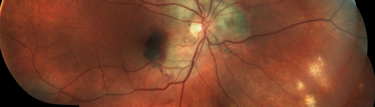
During the primary infection, the fungus enters the bloodstream and travels to the other parts of the body, where small areas of inflammation form. These areas of inflammation can also occur under the retina, where they are painless and go unnoticed. When the inflammation goes away, a scar is formed. Ten to thirty years later, these scar areas may result in the growth of abnormal blood vessels under the retina. As the blood vessels grow into this area, they can ooze serous (clear) fluid and blood. If this occurs in the peripheral retina, patients are unlikely to notice any changes to their vision. Unfortunately, most of the time, this occurs near the macula, where it can greatly affect central vision.
If left untreated, this leakage can cause permanent loss of central vision, but if detected early, laser eye surgery can often stop the leaking and preserve central vision. If this leaking has occurred into the macula, patients will notice distorted vision, and objects may appear smaller with that eye.
Treating Ocular Histoplasmosis
After diagnosing Ocular Histoplasmosis, your ophthalmologist will need to pinpoint where the leaking, abnormal blood vessels are. This is done by performing a test called a fluorescein angiogram. This test is done by injecting vegetable dye into an arm vein. From there, the dye circulates to the retina at the back of your eye, where photographs are taken with a special camera to study the retinal blood vessel supply and to determine if abnormal blood vessels are present.
Abnormal blood vessels can be treated with laser surgery, but sometimes, the abnormal blood vessels are too advanced. Whether or not laser surgery is performed is based on the location of the leaking and whether laser treatment is more likely to prevent severe loss of vision than if no laser treatment is done.
If laser treatment is successful, you can expect a significant decrease in distortion, but vision may not improve significantly. The main goal of the laser is to prevent more severe loss of central vision that usually occurs if the leaking blood vessels continue to grow and bleed.
After the laser treatment, a scar forms where the laser beam comes in contact with the retina. This creates a blind spot just off to the side of central vision. This permanent blind spot is a trade-off in order to save the remaining portions of central vision.
Even when laser treatment is successful, new leaking blood vessels can reappear at a later time, leading to further visual damage. Because of this, it is essential to check vision daily with an Amsler grid and report any changes immediately. Despite the possibility of central vision loss, the good news is that peripheral or side vision is almost always maintained, so mobility is not affected. In general, people do not go “blind,” even in the most severe cases. There are also several newer treatments available, including photodynamic therapy and several medications that can be injected into the eye to help stop abnormal blood vessels from leaking.