Branch Retinal Vein Occlusion
A branch retinal vein occlusion is an obstructed or blocked vein in the retina at the back of the eye. Arteries bring fresh nutrients and oxygen-rich blood to the tissues. Veins drain away that oxygen- and nutrient-depleted blood. While blood flow into the eye is important, blood flow out of the eye is equally important to ensure circulation. If this outflow from the eye is blocked, which is what happens with a retinal vein occlusion, the blood has nowhere place to go and “backs up,” rupturing the blood vessel walls. Without a way for the blood to flow out of the eye, blood overflows on and into the retina. Similar to the swelling that occurs when a tourniquet is kept in place on an arm too long, swelling occurs in the retina.
How much vision loss occurs will depend on how close the blood vessel blockage is to the center of vision (the macula) and if the blockage is partial or complete. Branch retinal occlusions may cause significant damage to vision. Depending on the duration and extent of the blockage, your vision may improve, worsen, or remain the same.
Vein occlusions can occasionally be complicated by abnormal blood vessel growth, which can later result in bleeding into the vitreous jelly of the eye or severe glaucoma. Careful follow-up examinations will monitor and allow early detection of these uncommon complications.
Retinal Vein Occlusion Treatment in Atlanta
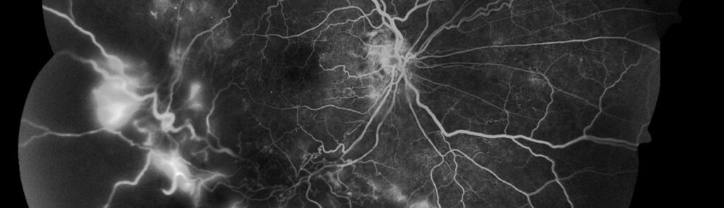
A special in-office test called a fluorescein angiogram or an OCT (optical coherence tomography) may be used to help confirm the diagnosis or guide treatment. The test is valuable because it will demonstrate the degree of diseased retina and can even reveal hidden damage, providing critical information for both treatment and predicting visual recovery.
Fortunately, there are treatments for branch retinal vein occlusions. If your vision does not improve significantly, or if your vision remains poor, laser eye surgery may be recommended. Laser treatments almost double the chance for visual improvement if there is macular edema.
Another treatment for macular edema is the injection of medications into the eye, like steroids or vascular endothelial growth factor inhibitors. If there are more serious cases where abnormal vessel growth occurs, a more extensive laser treatment may be utilized. In rare eyes with bleeding into the vitreous (jelly inside the eye) that fail to respond to conventional treatment, then vitreous surgery to remove the blood in the jelly of the eye becomes an option. This is inpatient surgery that may require a hospital stay. It is often combined with laser treatment.
Certain medical conditions increase the risk of developing these blood vessel blockages. The most common include hypertension (high blood pressure), diabetes, or atherosclerosis (hardening of the arteries, and especially carotid artery narrowing). Being referred to your primary care physician for reevaluation of these conditions may be valuable. Recognition and control of these medical conditions may prevent these blood vessel blockages from recurring in this eye, occurring in the other eye, or elsewhere in the body.